

Global Automated Microscopy Market Size and Forecast – 2026 To 2033
The global automated microscopy market is expected to grow from USD 8.72 Bn in 2026 to USD 14.08 Bn by 2033, registering a compound annual growth rate (CAGR) of 7.1% from 2026 to 2033. The market for global automated microscopy is poised for significant expansion, fueled by the rising government investment in biomedical research and advanced imaging technologies.
The U.S. National Institutes of Health (NIH) awarded USD 35.3 billion in competing and noncompeting grants for biomedical research in FY2025, while its FY2026 program-level funding reached USD 47.5 billion, supporting continued investment in research infrastructure and next-generation technologies.
Key Takeaways of the Global Automated Microscopy Market
- Optical microscopes are projected to hold 48.6 % of the global automated microscopy market share in 2026, making it dominant product type segment, across Europe supported by the region’s established pathology and microscopy infrastructure and regulatory framework for digital imaging technologies. The European Commission’s Medical Device Coordination Group (MDCG) issued updated guidance in 2025 on the qualification and classification of medical-device software under the EU MDR/IVDR, supporting regulatory pathways for digital imaging and pathology technologies.
- Automated image acquisition is projected to hold 35.2 % of the global automated microscopy market share in 2026, making it dominant automation type segment, with Asia Pacific gaining traction from increasing government-led procurement of automated imaging systems for biomedical research. For instance, in April 2025, India’s ICMR-National Institute of Virology invited quotations for an inverted fluorescence-based automated live-cell imaging system, requiring automated imaging capabilities for biological research applications.
- Life sciences research is projected to hold 31.8 % of the global automated microscopy market share in 2026, making it dominant application segment, across Asia Pacific supported by expanding government-backed microscopy infrastructure and biological imaging initiatives. For instance, India’s Department of Biotechnology operates the Indian Biological Images Archive (IBIA), which includes microscopy, confocal, scanning electron, light-sheet, and histopathology images to support biological research and AI-based image analysis.
- North America market maintains dominance with an expected share of 34.7 % in 2026, bolstered by strong government-backed investments in advanced microscopy infrastructure and biomedical research. In May 2026, the U.S. National Institutes of Health (NIH) awarded USD 425,494 to the University of North Carolina at Chapel Hill to acquire a high-speed light-sheet microscope for neuroscience, cancer biology, immunology, and therapeutics research.
- Asia Pacific is expected to exhibit the fastest growth in the global automated microscopy market, registering an estimated CAGR of 9.0 % during 2026–2033, driven by increasing government support for indigenous automated imaging and AI-enabled diagnostics. For instance, in February 2026, India’s Technology Development Board (TDB), under the Department of Science & Technology, supported the development and commercialization of “Autoscope,” a fully automated whole-slide imaging and AI-driven diagnostic system.
Segmental Insights
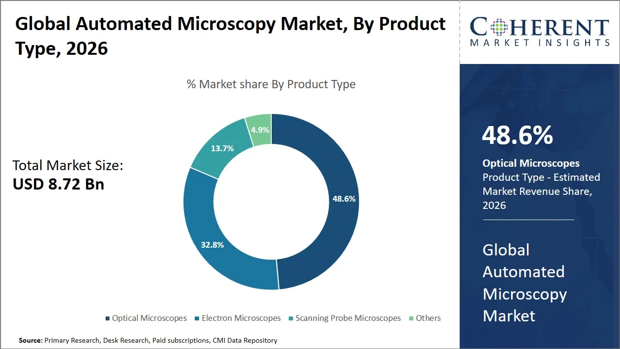
Why Do Optical Microscopes Dominate the Global Automated Microscopy Market?
Optical microscopes are projected to hold the market share of 48.6% in 2026, due to its multi-functionality, ease in sample preparation and ease to application in biological, clinical and materials studies. Also, optical microscopy can be integrated along with other imaging applications such as fluorescence, confocal and high content imaging with automated acquisition, focusing and image-analysis workflows. High throughput imaging and demand for reproducible results accelerate the uptake in optical microscopy.
For instance, in January 2026, The University of Huddersfield entered a contract to purchase a high-content imaging microscope system with automated acquisition across multi-well plates, slides and dishes, confocal microscopy, and high-throughoutput image processing functions. The procurement demonstrates persistent institutional demand for advanced automated optical microscopy for research applications. (Source: European Union)
- Current Industry Events of 2026
- Market Size Estimation
- Regional Breakdown
- Competitive Landscape
- Customer Intelligence
- Segmental Analysis
- Pricing Analysis
- Key Market Drivers, Challenges & Future Trends
- Customized Insights Section
Why Does Automated Image Acquisition Represent the Largest Automation Type Segment in the Automated Microscopy Market?
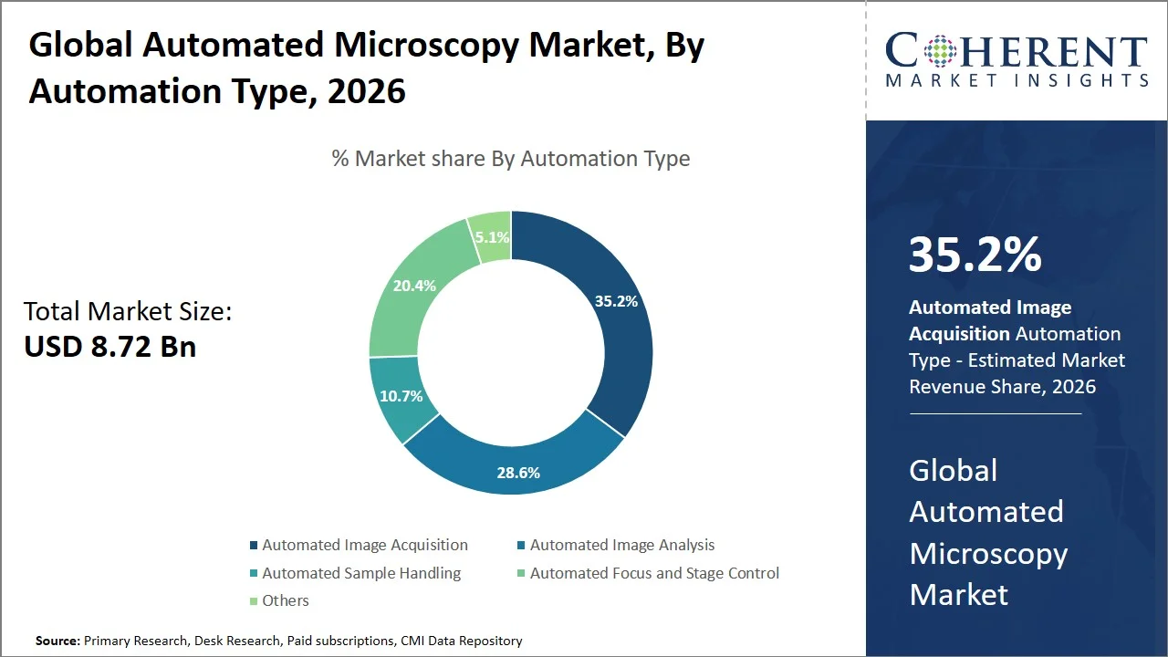
Automated image acquisition is projected to hold a market share of 35.2% in 2026, due to its ability to achieve repeatable results when a microscope is used to collect an image at a number of sample sites, over several slides without direct human input. Automated stages and focusing, programmable image acquisition, and auto-exposure provide increased throughput, reliability, and precision useful for throughput biologics and materials microscopy. This allows microscope use to transition toward automated, analysis-based workflows through integration of automated image acquisition with automated analysis.
For instance, in July 2026, the U.S National Institute of Standards and Technology (NIST) initiated procurement of an automated inverted microscope that have computer-controlled focus, computer-controlled objective and filter change, computer-controlled scan stage, and computer programs to control both the microscope, and automated image acquisition. The system is intended to support NIST’s organ-on-a-chip and micro physiological-systems research.
Life Sciences Research Segment Dominates the Global Automated Microscopy Market
The life sciences research segment is projected to hold a market share of 31.8% in 2026, driven by increased demand from life science research for automating cell/tissue imaging or visualizing biological samples in high-throughput to ensure reproducibility. Automated microscopy offers real-time observation capabilities for dynamic imaging of living cells, quantitative cellular parameters, and easy handling of large biological datasets without manual handling. Increasing applications of live cell imaging, cellular phenotyping, and advanced image processing further contribute to growth in life sciences. Increased combination of AI driven analysis and automation further helps in scaling out and efficient application of microscopy-based research.
For instance, in February 2026, Central University of Tamil Nadu initiated the process of purchasing laboratory instruments to set-up a Live Cell Imaging Facility as part of an ongoing Department of Science & Technology (DST) PURSE project to further enhance live-cell imaging facility for various biological disciplines across all departments to promote live life sciences research. (Source: National Informatics Centre)
Current Events and their Impact
Current Events | Description and its Impact |
Canada Funded Advanced Microscopy Infrastructure (August 2026) |
|
European Commission Advanced Microscopy Research Funding (May 2026) |
|
UK Government Procurement Expanded Automated Microscopy Capacity (March 2025) |
|
Automated Microscopy Market Dynamics
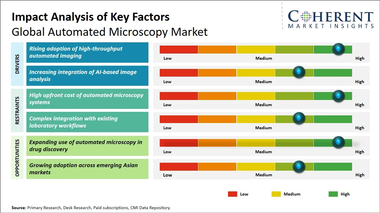
Market Drivers
- Rising adoption of high-throughput automated imaging: The growing need to process large numbers of biological samples rapidly and consistently is increasing the adoption of high-throughput automated imaging systems. Automated whole-slide scanning, batch processing, and quantitative image analysis enable laboratories to generate larger datasets while reducing manual imaging time and operator variability. This trend is particularly relevant to spatial biology, biomarker discovery, drug research, and translational studies. For instance, in August 2026, the U.S. National Institutes of Health awarded USD 444,159 to the University of Wisconsin System for an automated quantitative pathology imaging and analysis system capable of high-throughput whole-slide, tissue-array, and multispectral imaging. The system is intended to support spatial biology, biomarker discovery, and drug-discovery research.
- Increasing integration of AI-based image analysis: The increasing complexity and volume of microscopy data is accelerating the integration of AI-based image analysis into automated microscopy workflows. Machine learning can automate image interpretation, object recognition, segmentation, and quantitative analysis, enabling researchers to extract biological and material insights with less manual intervention. This is particularly important as microscopy increasingly generates high-dimensional datasets that require scalable computational analysis. For instance, in July 2026, the U.S. Department of Energy awarded USD 1.1 million through its SBIR program to develop an open analysis platform incorporating machine vision and AI for electron microscopy. The project focuses on automated image evaluation, object tracking, feedback control, and optimization of microscopy parameters, directly advancing AI-enabled microscopy workflows.
- Increasing government investment in advanced microscopy infrastructure: Growing government investment in advanced research infrastructure is encouraging laboratories and academic institutions to upgrade microscopy capabilities with automated imaging, motorized control, and integrated analysis. Public funding and institutional procurement are increasingly focused on high-end imaging systems that can support large-scale biological and materials research, creating opportunities for automated microscopy adoption. For instance, in August 2026, India’s Department of Higher Education invited bids through the Government e-Marketplace for a high-content automated confocal imaging, screening, and analysis system at the University of Delhi. The procurement directly supports automated high-throughput imaging and analysis for biological research.
Emerging Trends
- Integration of AI and machine learning in microscopy: Automated microscopy systems are increasingly incorporating AI-based image segmentation, classification, feature extraction, and phenotype recognition, enabling faster and more consistent interpretation of complex image datasets.
- Expansion of 3D and high-content imaging: Growing use of organoids, spheroids, 3D cell cultures, and complex biological models is increasing demand for automated microscopy capable of high-throughput, multidimensional imaging and quantitative analysis.
- Shift toward integrated laboratory automation: Microscopy is increasingly being integrated with robotic sample handling, automated stages, environmental controls, image management, and laboratory information systems, creating end-to-end automated workflows with reduced manual intervention.
Regional Insights
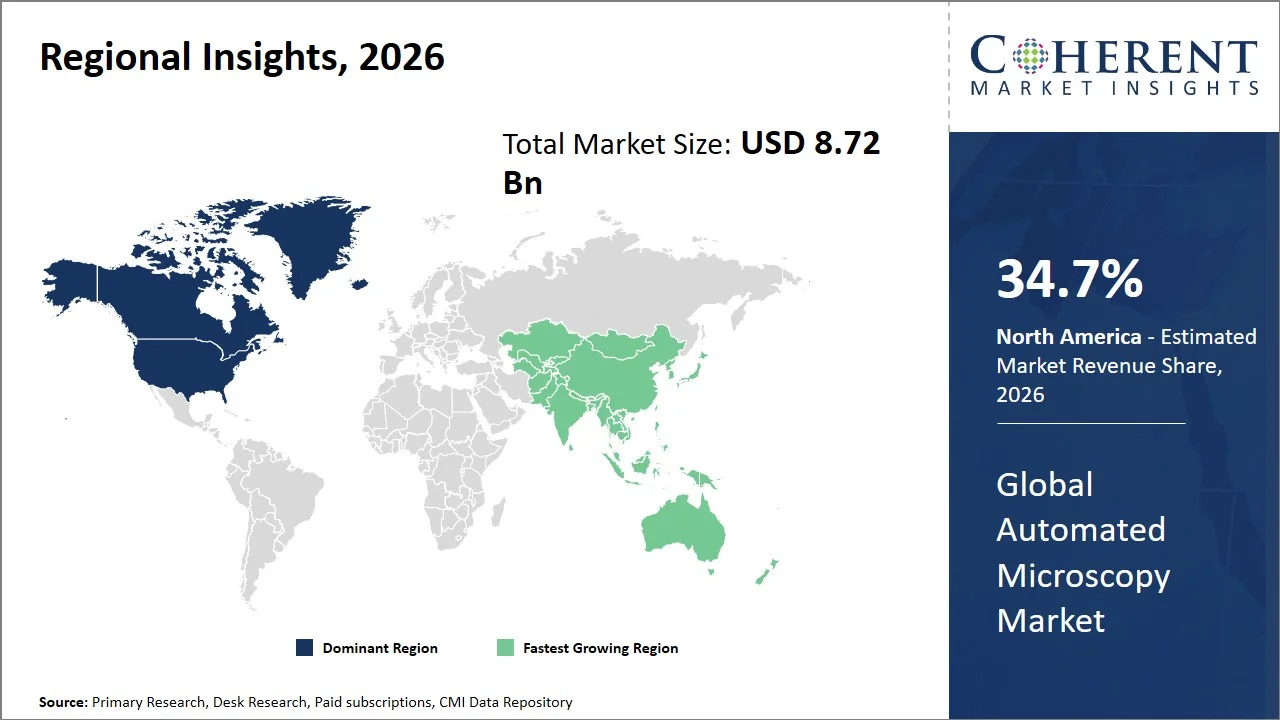
Why is North America a Strong Market for Automated Microscopy?
North America leads the global automated microscopy market, accounting for an estimated 34.7% share in 2026, attributed to the region’s robust healthcare systems, well-established research infrastructure, high acceptance of lab automation technology, and concentrated research & development in the biomedical as well as industrial sectors. The existence of prominent academic, pharmaceutical, and research segments encourages an increased demand for highly productive imaging techniques and AI-enabled microscope technologies within the region.
In addition, supportive initiatives from the governments and leading agencies (such as the NIH) stimulate technical improvements, contributing to the innovations for future imaging modalities. For instance, in February 2026, the U.S. National Institutes of Health (NIH) Common Fund highlighted its Advancing Non-Invasive Optical Imaging Approaches for Biological Systems (NIOI) endeavor, it develops next-generation optical imaging technologies to acquire high resolution information from deeper live biological systems.
Why Does Asia Pacific Automated Microscopy Market Exhibit High Growth?
Asia Pacific is expected to exhibit the fastest growth in the global automated microscopy market, registering an estimated CAGR of 9.0% during 2026–2033. The region is projected to account for approximately 25.6% of the global market in 2026, fueled by increased healthcare spending, expanding R&D efforts in biopharma, and the up gradation of lab infrastructure. Demand for more high-throughput and repeatable microscopy workflows is intensifying with high adoption of automated imaging and AI driven analysis technologies.
Government initiatives in countries such as China, Japan, and India aimed at fostering innovation, combined with the booming CRO industry, expanding pharmaceutical & life science, and advancements in digital health is widening the scope of market expansion. For instance, in August, 2026, the government procurement program was launched by the Peking University for a 3D light-sheet microscopy system valued USD 0.552 million (RMB 3.7 million) that applies in 3D fluorescence imaging of rapid whole organs, mice cleared tissues and whole human samples. (Source: Ministry of Finance of the People's Republic of China)
Global Automated Microscopy Market Outlook for Key Countries
Why is the U.S. Leading Innovation and Adoption in the Automated Microscopy Market?
The U.S. has been in the forefront of leading innovation and adoption in the automated microscopy market being spurred through combination of AI, robotics and microscopy to create fully autonomous research workflows across biomedical sciences and materials science. The U.S system will shift from automated image acquisition toward images, which can analyze them and adapt imaging parameters, and direct experiments in real time. This will further increase demand for intelligent microscopy systems that can facilitate high-throughput data-based analysis and minimize operator intervention.
Is Japan a Favorable Market for Automated Microscopy Market?
Japan has proven to be a strong market opportunity, particularly in AI microscopy, life sciences automation and digital pathology. There is strong demand driven by government supported programs that are accelerating the deployment of AI, robots, and automated experimentation into the overall research infrastructure, which necessitates the need for cutting-edge microscopes and imaging analysis. Additionally, the country is working to mature its digital/AI medical technology review processes which will allow for broader use of automated imaging in a clinical setting.
Is China Emerging as a Key Growth Hub for the Automated Microscopy Market?
China is likely to be a major growth center, with high potential specifically for AI enabled electron microscopy and automated materials characterization. Trends toward autonomous microscopes where a connection is made between the sample handling, imaging and analysis, rather than independent automated functions, are forming the foundation of future markets. China’s superiority in advanced materials, chemistry and high-end scientific instrumentation creates a broad application base for these next-generation microscopy platforms.
Why Does Germany Top the European Automated Microscopy Market?
Germany constitutes a significant and mature market for automated microscopy attributable to country's robust position in the precision engineering, sophisticated optical technologies and industrial microscopy. The country has well-established semiconductor industry, automotive industry and materials industry that will maintain demand for automated inspection, automated dimensional measurement and high-resolution imaging. The strong research community in the country will also boost adoption of advanced automated microscopy systems.
Is Automated Microscopy Market Developing in India?
As India moves on a trajectory of growth into an emerging economy, both increased availability of shared cutting-edge microscopy capabilities and deployment of AI for biological images will help build momentum for automating microscopy in India’s research institutions, academia, and industry. Government supported platforms such as DBT-SAHAJ is democratizing access to state-of-the-art optical and electron microscopy in research institutions and the country is developing AI powered biological imaging capabilities. Together these will provide a firm backbone for proliferation of automated microscopy in life sciences, diagnostics and drug discovery.
Application of Automated Microscopy in Drug Discovery and High-Content Screening
Application Area | Key Use of Automated Microscopy | Primary Output |
Primary Screening | Automated imaging of large compound libraries | Hit identification |
Phenotypic Screening | Multiparametric cellular imaging | Cellular response profiles |
Toxicity Screening | Automated detection of cellular toxicity | Toxicity assessment |
Compound Profiling | High-content imaging of cellular phenotypes | Compound characterization |
Target Validation | Quantitative imaging of target-related cellular changes | Target confirmation |
How is the expanding use of automated microscopy in drug discovery creating new growth opportunities in the automated microscopy market?
The widespread usage of automated microscopy in drug discovery processes is driving considerable opportunities for vendors as the technology can test a multitude of drug candidates as well as cellular responses with a help of high-content imaging. Automated imaging facilitates an increased screening throughput; the efficiency and reproducibility of screening being enhanced by the minimized amount of researcher involvement and a more quantitative phenotypic analysis being conducted. The integration of microscopy with computational analysis additionally aids in the more rapid identification of potential therapeutic candidates. For instance, in April 2026, the U.S. National Center for Advancing Translational Sciences (NCATS) granted USD 436,250 to the Broad Institute to develop a drug-repurposing pooled image-based system named RAPID, which will screen 26,400 drug-disease compounds using high-dimensional image profiling and prove the immediate usability of automated microscopy-based images in drug discovery.
Market Players, Key Development, and Competitive Landscape
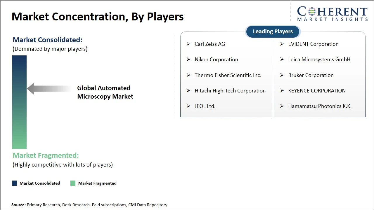
Key Developments
- In August 2026, Aliko Scientific signed new distribution agreements in Romania and Saudi Arabia, covering its microscopy, imaging, FISH, and liquid-based cytology solutions. The agreements strengthen the company’s international distribution network and expand access to automated microscopy and imaging technologies in oncology diagnostics.
- In May 2026, Nikon Corporation launched the ECLIPSE LV100AMS, a one-click automated microscope designed for industrial inspection and quality control. The system integrates automated image acquisition, motorized control, and AI-powered image analysis to streamline inspection workflows. It enables faster, standardized inspections while reducing operator-dependent variability and human error.
- In March 2025, Thermo Fisher Scientific Inc. introduced the Vulcan Automated Lab, an AI-powered automated workflow integrating robotic handling and electron microscopy for semiconductor analysis. The solution enables high-volume TEM metrology while reducing operator burden and improving laboratory productivity.
Competitive Landscape
The global automated microscopy market is moderately competitive, with market dynamics shaped by advancements in automated imaging, AI-enabled image analysis, high-throughput microscopy, and integrated laboratory workflows. Market participants are increasingly focusing on improving imaging speed, resolution, reproducibility, analytical accuracy, and workflow automation while expanding applications across life sciences research, drug discovery, clinical diagnostics, materials science, and industrial inspection. Competition is also shifting toward integrated microscopy platforms that combine automated acquisition, focus and stage control, sample handling, and image analysis capabilities. Key focus areas include:
- Development of AI-enabled image analysis and automated image interpretation
- Advancement of high-throughput and high-content imaging platforms
- Integration of automated image acquisition, autofocus, and stage control
- Expansion of automated sample handling and end-to-end microscopy workflows
- Development of integrated microscopy hardware, software, and data-analysis solutions
Market Report Scope
Automated Microscopy Market Report Coverage
| Report Coverage | Details | ||
|---|---|---|---|
| Base Year: | 2025 | Market Size in 2026: | USD 8.72 Bn |
| Historical Data for: | 2020 To 2024 | Forecast Period: | 2026 To 2033 |
| Forecast Period 2026 to 2033 CAGR: | 7.1% | 2033 Value Projection: | USD 14.08 Bn |
| Geographies covered: |
| ||
| Segments covered: |
| ||
| Companies covered: | Carl Zeiss AG, EVIDENT Corporation, Nikon Corporation, Leica Microsystems GmbH, Thermo Fisher Scientific Inc., Bruker Corporation, Hitachi High-Tech Corporation, KEYENCE CORPORATION, JEOL Ltd., Hamamatsu Photonics K.K. | ||
| Growth Drivers: |
| ||
| Restraints & Challenges: |
| ||
Analyst Opinion (Expert Opinion)
- In the coming years, global automated microscopy market is expected to evolve toward intelligent, interconnected, and increasingly autonomous imaging platforms. Future systems are likely to combine AI-based image interpretation with automated acquisition, focusing, stage movement, sample handling, and real-time decision-making. The shift from microscopy as an imaging instrument toward a data-driven analytical workflow is expected to become a major competitive direction, particularly in life sciences and drug discovery.
- The maximum opportunities are foreseen within optical microscopes for drug discovery and development in China. Optical systems offer broad applicability for cellular and phenotypic imaging, while drug discovery increasingly depends on high-throughput and multiparametric assessment of cellular responses. China presents an attractive opportunity because of its expanding pharmaceutical research base, increasing investment in laboratory automation, and growing demand for advanced imaging capabilities.
- In order to gain a competitive advantage, market players should move beyond hardware differentiation and develop integrated microscopy ecosystems combining imaging, automation, AI analysis, and workflow management. Priority should be given to application-specific solutions that deliver measurable improvements in throughput, reproducibility, and analytical accuracy. Establishing strong software capabilities, interoperability with laboratory systems, and localized application support can further strengthen customer retention and competitive positioning.
Speak to the analyst
Want personalized insights?
Take the findings above to one of our principal consultants, or have the report rebuilt around the segments, geographies and competitors you actually track.
Market Segmentation
- Product Type Insights (Revenue, USD Bn, 2021 - 2033)
- Optical Microscopes
- Electron Microscopes
- Scanning Probe Microscopes
- Others
- Automation Type Insights (Revenue, USD Bn, 2021 - 2033)
- Automated Image Acquisition
- Automated Image Analysis
- Automated Sample Handling
- Automated Focus and Stage Control
- Others
- Application Insights (Revenue, USD Bn, 2021 - 2033)
- Life Sciences Research
- Drug Discovery and Development
- Clinical Diagnostics
- Materials Science
- Industrial Inspection
- Others
- End User Insights (Revenue, USD Bn, 2021 - 2033)
- Pharmaceutical and Biotechnology Companies
- Hospitals and Diagnostic Laboratories
- Academic and Research Institutions
- Contract Research Organizations
- Industrial Companies
- Others
- Regional Insights (Revenue, USD Bn, 2021 - 2033)
- North America
- U.S.
- Canada
- Latin America
- Brazil
- Argentina
- Mexico
- Rest of Latin America
- Europe
- Germany
- U.K.
- Spain
- France
- Italy
- Russia
- Rest of Europe
- Asia Pacific
- China
- India
- Japan
- Australia
- South Korea
- ASEAN
- Rest of Asia Pacific
- Middle East
- GCC Countries
- Israel
- Rest of Middle East
- Africa
- South Africa
- North Africa
- Central Africa
- North America
- Key Players Insights
- Carl Zeiss AG
- EVIDENT Corporation
- Nikon Corporation
- Leica Microsystems GmbH
- Thermo Fisher Scientific Inc.
- Bruker Corporation
- Hitachi High-Tech Corporation
- KEYENCE CORPORATION
- JEOL Ltd.
- Hamamatsu Photonics K.K.
Sources
Primary Research Interviews
- Automated microscopy system developers and microscopy automation specialists
- Biomedical imaging and image-analysis scientists involved in automated microscopy workflows
- Pharmaceutical and biotechnology R&D scientists using automated microscopy for drug discovery and high-content screening
- Clinical laboratory and digital pathology specialists using automated imaging systems
- Materials science and semiconductor inspection specialists using automated microscopy
Stakeholders
- Pharmaceutical and biotechnology companies using automated microscopy for drug discovery and life sciences research
- Hospitals, diagnostic laboratories, and pathology centers adopting automated imaging workflows
- Academic and research institutions conducting cell biology, materials science, and microscopy research
- Contract research organizations providing high-content imaging and screening services
- Industrial and semiconductor manufacturers using automated microscopy for inspection and quality control
- End-use Sectors
- Pharmaceutical and biotechnology companies
- Contract research organizations (CROs)
- Academic and research institutions
- Hospitals and diagnostic laboratories
- Clinical and digital pathology laboratories
- Industrial inspection and quality-control organizations
- Semiconductor and electronics manufacturers
- Materials science and nanotechnology research organizations
- Biotechnology and life-sciences research laboratories
- Regulatory & Health Bodies
- U.S. Food and Drug Administration (FDA) – medical imaging, digital pathology, and diagnostic device regulation
- European Medicines Agency (EMA) – medicinal-product and clinical research regulatory information
- Medicines and Healthcare products Regulatory Agency (MHRA), U.K. – medical-device and diagnostic technology regulation
- Pharmaceuticals and Medical Devices Agency (PMDA), Japan – medical-device and diagnostic product regulation
- National Medical Products Administration (NMPA), China – medical-device and diagnostic technology regulation
Databases
- ClinicalTrials.gov – clinical studies involving imaging, diagnostics, and drug development
- BioImage Archive – publicly accessible biological imaging datasets
- NIH RePORTER – publicly funded biomedical imaging and microscopy research
- Materials Data Facility – materials characterization and microscopy-related research datasets
Journals
- Nature Methods – advanced microscopy, imaging, and quantitative image analysis
- Microscopy and Microanalysis – microscopy technologies, imaging methods, and analytical microscopy
- Journal of Microscopy – optical, electron, scanning probe, and computational microscopy
- Cytometry Part A – quantitative cell analysis, imaging, and high-content cellular analysis
- Journal of Cell Science – cellular imaging, microscopy, and cell biology research
Associations
- Microscopy Society of America (MSA)
- Royal Microscopical Society (RMS)
- International Federation of Societies for Microscopy (IFSM)
- Society for Imaging Informatics in Medicine (SIIM)
- SPIE – The International Society for Optics and Photonics
Public Domain Sources
- U.S. National Institutes of Health (NIH) – biomedical imaging and microscopy research funding
- National Institute of Biomedical Imaging and Bioengineering (NIBIB) – imaging technology and biomedical engineering research
- U.S. Food and Drug Administration (FDA) – digital pathology and medical imaging device information
- National Institute of Standards and Technology (NIST) – microscopy, measurement science, and imaging standards
- European Commission – Horizon Europe funding for advanced imaging and life-sciences research
- Department of Biotechnology (DBT), India – biotechnology, biological imaging, and research infrastructure initiatives
Proprietary Elements
- CMI Data Analytics Tool, Proprietary CMI Existing Repository of information for last 10 years.
Get access to 250+ pages report
Every segment, forecast, competitor profile and data table behind the analysis above — available for instant download.
- ESOMAR Member
- ISO 9001:2015 Certified
- D-U-N-S Registered
- GDPR & CCPA Compliant
Missing comfort of reading report in your local language? Find your preferred language:
Frequently Asked Questions
The global automated microscopy market is estimated to be valued at USD 8.72 Bn in 2026 and is expected to reach USD 14.08 Bn by 2033.
Optical microscopes dominate due to their broad use in life sciences research, drug discovery, clinical diagnostics, and industrial inspection, along with their compatibility with automated imaging and analysis workflows.
Automated microscopy is the use of computer-controlled imaging, focusing, stage movement, sample handling, and image analysis to perform microscopic examination with minimal manual intervention.
Automated microscopy uses computer-controlled imaging, focusing, stage movement, sample handling, and software-based analysis to automate microscopic examination.
Key benefits include higher throughput, improved reproducibility, reduced manual intervention, faster analysis, and consistent image quality.
AI enables automated image segmentation, object detection, classification, feature extraction, and quantitative analysis of complex microscopy images.
High-content imaging combines automated microscopy with multiparametric image analysis to measure multiple cellular characteristics across large sample sets.